Researchers in Spain, the United Kingdom and Germany have demonstrated that graphene electrodes can capture the sluggish electrical disturbances that ripple through brain tissue after an ischaemic stroke, opening a route to monitoring damage as it unfolds rather than after the fact. The work, published in the journal Brain, was carried out by Spain's Consejo Superior de Investigaciones Científicas (CSIC), the University of Manchester, the Catalan Institute of Nanoscience and Nanotechnology (ICN2) and the German instrumentation firm Multi Channel Systems.
An ischaemic stroke occurs when a clot cuts blood flow to part of the brain. The initial lesion is only the beginning: waves of disrupted electrical activity, known as spreading cortical depolarisations, travel outward from the core of the injury and can push surrounding tissue toward irreversible damage. Clinicians have long wanted to intercept those waves, but conventional electrodes struggle to resolve the very slow, low-frequency signals involved.
Why graphene changes the picture
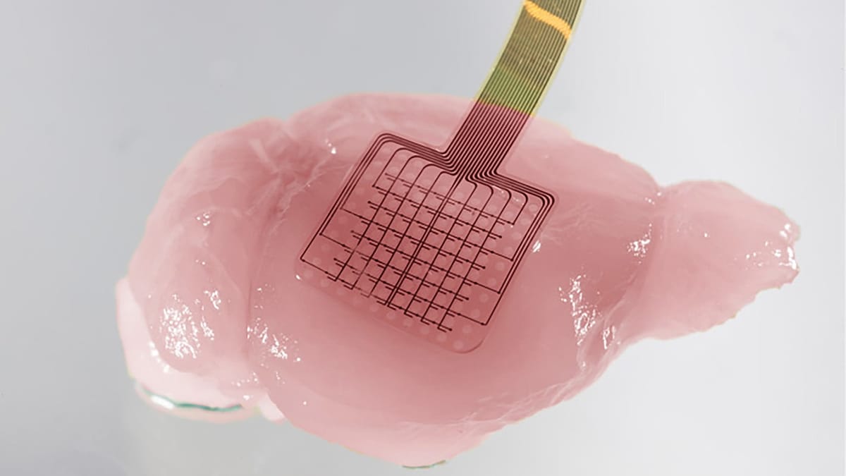
Graphene's combination of high conductivity and sensitivity to tiny electric fields makes it unusually well suited to recording slow cortical events. Placed directly on the brains of mice, the team's sensors picked up the depolarisation waves in far finer detail than standard arrays. The signal signatures, the researchers found, allowed them to sort tissue into three categories: relatively healthy, at risk, and already severely compromised.
The recordings also revealed a predictive relationship with blood flow. In healthier regions, perfusion rose as the brain tried to sustain recovery. In vulnerable zones, it could fall further, deepening the injury. That link suggests the electrical waves are not merely a symptom of damage but an active driver of it.
"In broad terms, the study shows how cutting-edge technologies can not only improve the way brain activity is measured, but also reveal new and important information about how brain lesions develop and how they might be treated," said Rob Wykes, a researcher at the University of Manchester.
The team went further and tested a low dose of ketamine, a drug already in clinical use as an anaesthetic and for treatment-resistant depression. In the animal models, ketamine shortened the duration of the harmful waves, improved the blood-flow response and reduced the overall extent of brain damage. The finding points to a possible neuroprotective role for the drug after stroke, though the authors are careful to note that no equivalent benefit has yet been shown in humans.
A decade of sensor development
The result is the product of a long-running collaboration between Spanish and British institutions aimed at building graphene devices capable of recording brain signals. Anton Guimerà-Brunet, a CSIC researcher at the Institute of Microelectronics of Barcelona, noted that low-frequency brain activity remains difficult for existing tools to capture. "We have been developing our graphene-based technology for years, with our first results published in 2018, and the challenge has been to standardise and refine it for use in relevant preclinical studies," he said.
The broader context is a European research landscape that has become notably strong in neurotechnology, from Barcelona's microelectronics cluster to Manchester's graphene expertise — the material was first isolated at the university in 2004. Similar momentum is visible elsewhere on the continent, including Greek work on Alzheimer's models and studies of how the brain manages memory, both of which reflect the same push to translate basic neuroscience into clinical tools.
For now, the graphene platform remains at the preclinical stage. The path to human use will require larger animal studies, regulatory clearance and devices that can be deployed safely in an acute stroke setting, where minutes matter and imaging suites are already crowded.
What it could mean for patients
If the technology translates, its most immediate value may be diagnostic rather than therapeutic. Real-time monitoring of brain tissue during a stroke could help clinicians identify which patients are most likely to suffer secondary injury and adjust treatment accordingly — for instance, by intensifying blood-pressure management or selecting candidates for neuroprotective drugs.
The ketamine finding, meanwhile, adds to a small but growing body of evidence that the drug's effects extend beyond anaesthesia. Any stroke application would need careful dose-finding and safety work, since ketamine's psychotropic effects and its use in emergency settings raise practical questions.
What the study does establish is a proof of principle: that the slow electrical storms following a stroke can be watched, classified and, in principle, interrupted. That is a meaningful step for a condition that remains one of Europe's leading causes of death and long-term disability, and where treatment options have barely changed in a generation.